
Anemia: The Symptoms, and the Three Ways the Body Runs Short of Red Cells
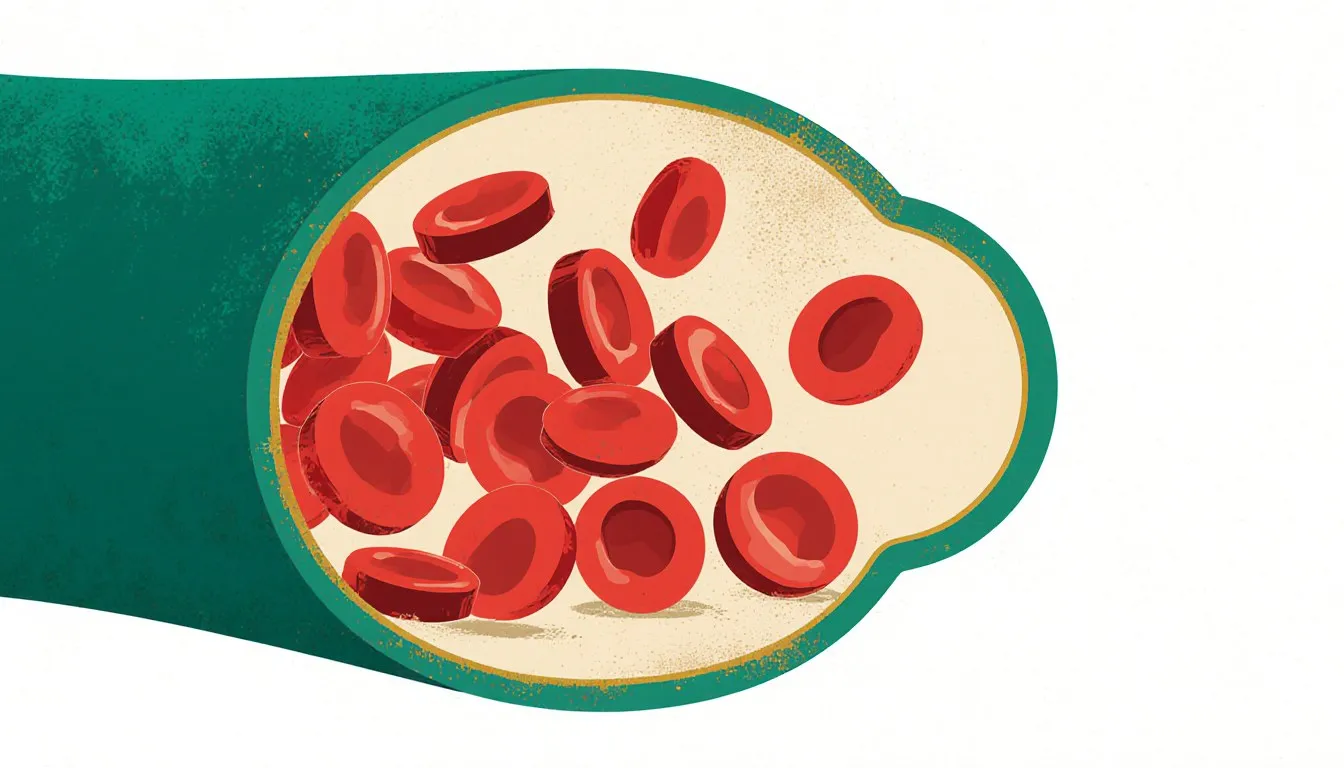
Anemia means the blood is delivering less oxygen than the body’s tissues are asking for. That one shortfall explains the whole condition: the tiredness that sleep does not fix, the paleness inside the lower eyelid, the breathlessness on a staircase that used to be unremarkable, and the heart rate that climbs to compensate. It also organizes every cause. There are only three ways the body can end up short of oxygen-carrying capacity. It can fail to make enough red blood cells, it can lose them, or it can destroy them faster than it replaces them. Iron deficiency, heavy periods, kidney disease, sickle cell disease, and a bleeding ulcer each belong in one of those three columns, and knowing which column a cause sits in is what decides how it gets investigated and how it gets treated.
This article explains a medical topic in general terms. It is not medical advice, it is not reviewed by a clinician, and it cannot account for your individual case. Decisions about your own diagnosis or treatment belong with your doctor.
What follows is the mechanism, the symptom picture it produces, the three families of cause, how the condition is found and sorted, what the standard treatments are, and what is still genuinely unsettled.
What is anemia, exactly?
Anemia is a shortfall in hemoglobin, the iron-containing protein inside red blood cells that binds oxygen in the lungs and releases it in tissue. A single hemoglobin molecule carries up to four oxygen molecules, and a red cell is packed with hundreds of millions of them. Fewer red cells, or less hemoglobin inside each one, means less oxygen arriving per heartbeat.
Red cells are consumable. They circulate for roughly 120 days, lose the ability to maintain their own membranes, and are cleared by the spleen and liver. The bone marrow replaces them continuously, at a rate usually given as around two million cells per second. That replacement line runs on a supply chain: iron for the heme group, vitamin B12 and folate for the DNA synthesis that lets precursor cells divide, erythropoietin from the kidneys as the signal to produce more, and a functioning marrow to do the work. Interrupt any link and production falls.
In practice, anemia is defined by hemoglobin concentration rather than by counting cells. Most laboratories and guidelines use thresholds that descend from World Health Organization definitions. The cutoff sits near 13 grams per deciliter in adult men. For non-pregnant adult women it sits near 12.
Those figures are not as fixed as they look on a printed report.
They were set from population data decades ago, and the reference ranges a given lab prints may differ slightly from them. Pregnancy uses lower cutoffs, and young children have values of their own. Those numbers are population conventions and have been revised as the evidence behind them has been re-examined. They are also adjusted for altitude and for smoking, both of which push hemoglobin up independently of anything being wrong.
One clarification worth making early, because it causes a lot of confusion in results portals. Hemoglobin is a concentration, so it moves when the fluid it is dissolved in moves. Pregnancy expands plasma volume substantially, which dilutes hemoglobin and can produce a low reading in someone whose total red cell mass is perfectly adequate. Endurance athletes show a milder version of the same effect. Real anemia and dilutional anemia look identical on the first line of a lab report and are not the same problem.
The word itself names a finding. It describes a state of the blood and says nothing about why the blood got that way. A result reading “anemia” opens the diagnostic question, and the rest of the workup exists to answer it. That is why this topic is organized around causes.
What does anemia feel like? The core symptoms
The common symptoms are the direct consequence of tissue receiving less oxygen, plus the body’s attempts to make up the difference. They cluster the same way across most types:
- Fatigue and weakness: the most common complaint and the least specific. Muscle and brain are the largest oxygen consumers, and both underperform first.
- Pallor: less hemoglobin means less of the pigment that gives blood its color. It shows most reliably in the conjunctiva, which is the lining inside the lower eyelid, the palmar creases, the nail beds, and the tongue, which is why clinicians look there rather than at the face. Judging pallor from overall skin tone is unreliable, and it is least reliable in people with darker skin, a limitation the examination literature has been explicit about for years.
- Shortness of breath on exertion: the lungs are working normally, and the blood leaving them is fully saturated. There is simply less carrier arriving to be loaded, so breathing harder does not fix the deficit.
- A fast or pounding heartbeat: the one lever the body can pull is circulating the available hemoglobin more often. Cardiac output rises, which produces palpitations and, over time, strain.
- Dizziness and lightheadedness, particularly on standing, and headache.
- Cold hands and feet: blood is preferentially shunted to the brain, heart, and kidneys, and the extremities are what gets sacrificed.
- Difficulty concentrating, often described as fogginess and often mistaken for stress or poor sleep.
Some symptoms point more specifically at cause. Iron deficiency has a distinctive set: restless legs, brittle or spoon-shaped nails, cracking at the corners of the mouth, a sore smooth tongue, hair shedding, and pica, the compulsion to chew non-food substances. The ice-craving version of pica, called pagophagia, is common enough in iron deficiency that its appearance in a history tends to get a clinician’s attention. Vitamin B12 deficiency can produce numbness, pins and needles, unsteady walking, and memory changes, because B12 is needed for the myelin sheath around nerves as well as for red cell production. Hemolysis, where red cells are destroyed early, can add jaundice and dark urine, since the breakdown of hemoglobin releases bilirubin faster than the liver clears it.
The honest caveat is that none of these are proof of anything on their own. Fatigue and breathlessness have dozens of causes, and a great many people with mild anemia notice nothing at all, which is precisely why so much of it is found on blood work drawn for an unrelated reason.
Do symptoms differ by how severe or how sudden the anemia is?
Yes, and the difference between slow and sudden is larger than the difference between mild and moderate.
When hemoglobin falls gradually over weeks or months, the body compensates, and it compensates well. Cardiac output increases. Blood flow is redistributed toward the organs that tolerate hypoxia worst. Inside the red cells, a molecule called 2,3-BPG rises, which shifts the oxygen dissociation curve so that hemoglobin releases its oxygen to tissue more readily instead of clinging to it. Plasma volume expands. The net effect is striking. People with long-standing anemia can reach hemoglobin levels that look alarming on paper and still walk into a clinic under their own power, describing nothing worse than being tired lately. Older adults in particular tend to attribute the whole picture to age.
Sudden loss behaves differently, because the problem is not only oxygen-carrying capacity but circulating volume. A significant acute hemorrhage produces a racing pulse, falling blood pressure, clammy skin, confusion, and collapse, on a timescale of minutes, with none of the compensation described above having had a chance to develop.
There is a laboratory trap in that scenario worth knowing about. Immediately after acute blood loss, whole blood is leaving the body, red cells and plasma together, so the hemoglobin concentration can read close to normal for a period. It falls as fluid shifts back into the vasculature and dilutes what remains. In an actively bleeding person, an early normal-looking number is an artifact of timing. Acute bleeding is assessed by clinical signs first and by the lab number second, for exactly this reason.
What causes anemia? The three underlying mechanisms
Red cell supply is a balance between production, retention, and survival. Anemia occurs when one of the three breaks down, and every named cause in the medical literature sorts into one of them.
- Too few red cells made: iron deficiency, vitamin B12 and folate deficiency, chronic kidney disease, chronic inflammatory illness, marrow disorders such as aplastic anemia and myelodysplastic syndromes, and marrow suppression from chemotherapy. The reticulocyte count is what places a case in this group. Reticulocytes are the newest red cells in the circulation, and when they are scarce in someone who is clearly anemic, the marrow is not replacing what is being used up, so the production line itself is where the fault sits. The sorting from there follows the supply chain. Cell size narrows the field before any other test does, ferritin and iron studies, B12 and folate levels, and kidney function each check one link in that chain, and when all of them come back unhelpful the question moves to the marrow itself, which a biopsy answers directly.
- Red cells lost: acute bleeding from trauma, surgery, childbirth, or a ruptured vessel, and chronic slow bleeding from heavy menstrual periods, ulcers, colon lesions, or intestinal parasites. Chronic loss presents as iron deficiency, because iron leaves the body with every millilitre of blood and the stores are drawn down faster than absorption can refill them. Sorting this group is largely a matter of locating the leak. A careful history accounts for most of it, specific questions about menstrual flow account for a good deal more, stool testing looks for blood the person would never have seen, and endoscopy inspects the surfaces where slow bleeding usually starts.
- Red cells destroyed early: inherited disorders of the red cell membrane, enzymes, or hemoglobin itself, and acquired destruction from autoimmune disease, certain drugs, mechanical damage, or infection. Here the reticulocyte count runs high, because the marrow is intact and working hard to replace cells that keep disappearing. Bilirubin rises, and so does LDH, or lactate dehydrogenase, an enzyme that spills into the blood when cells of any kind rupture. Haptoglobin falls, because that protein mops up hemoglobin released loose into the circulation and gets used up when red cells are breaking down in quantity. Sorting this group means establishing that destruction is happening and then identifying what is doing it. The smear shows the shape of the surviving cells, the hemolysis panel confirms that breakdown is under way, the direct antiglobulin test separates immune destruction from every other kind, and hemoglobin electrophoresis, which separates the different types of hemoglobin in a sample, allows an abnormal one to be named.
Two mechanisms often run together, and the combination is what makes real cases harder than the framework suggests. Someone with chronic kidney disease makes too little erythropoietin and may also be losing blood at dialysis. Someone with celiac disease absorbs iron poorly and may bleed from an inflamed gut. The framework sorts a workup. In a given person two of the three boxes are often ticked at once, and the second one is usually the one that gets missed.
Causes: not enough red blood cells being made
This is the largest of the three groups and the one that accounts for most anemia worldwide.
Iron deficiency
Iron deficiency is the single most common cause of anemia globally. Without enough iron, the marrow keeps dividing precursor cells but cannot fill them with hemoglobin, so the cells that emerge are small and pale. That is why iron deficiency shows up as a low mean corpuscular volume, the microcytic pattern on a blood count.
Iron reaches the body through a narrow gate. Absorption happens mainly in the duodenum, the first stretch of small intestine, and heme iron from meat is taken up more readily than the non-heme iron in plants, fortified grains, and supplements. The body has no route for excreting excess iron deliberately, so balance is regulated almost entirely at the point of absorption. Deficiency therefore arises from poor intake, poor absorption after conditions like celiac disease or bariatric surgery, raised demand in pregnancy and childhood growth, or, most often in adults, ongoing loss.
Vitamin B12 and folate deficiency
B12 and folate are required for DNA synthesis. When either runs short, precursor cells in the marrow grow but cannot divide on schedule, producing large, structurally abnormal cells: the macrocytic or megaloblastic pattern. Many of these cells die inside the marrow before they ever reach the circulation, a phenomenon called ineffective erythropoiesis.
B12 absorption depends on intrinsic factor, a protein made by the stomach lining. When an autoimmune process destroys the cells that produce it, the result is pernicious anemia, which was uniformly fatal before the 1920s and is now treatable. Low intake matters in strictly plant-based diets, since B12 is essentially absent from unfortified plant foods, and long-term use of certain medicines including metformin and proton pump inhibitors is associated with reduced absorption. Folate deficiency is more often dietary, and it also rises in pregnancy and in conditions with rapid cell turnover. The two deficiencies look nearly identical on a blood count, so both are tested. The reason matters. Giving folate alone can tidy up the blood picture in someone whose actual shortage is B12, while the nerve damage carries on underneath a normal-looking result.
Chronic kidney disease and chronic inflammation
The kidneys sense oxygen levels and release erythropoietin, the hormone that tells the marrow to increase production. As kidney function declines, that signal weakens, and anemia becomes an expected feature of advanced kidney disease.
Chronic inflammatory illness produces anemia by a different route, and it is one of the more elegant mechanisms in medicine. Inflammation raises a liver hormone called hepcidin, which shuts down the channel that moves iron out of storage cells and out of the gut lining. Iron is still present in the body but locked away from the marrow. Hepcidin evolved partly to starve invading bacteria of iron. So this is a defence system producing a side effect, and it explains why anemia accompanies rheumatoid arthritis, inflammatory bowel disease, chronic infection, and cancer. It also explains why ferritin, which rises with inflammation, is a slippery test in exactly these patients.
Bone marrow disorders
Less common, more serious. In aplastic anemia the marrow stops producing cells across all lineages, so red cells, white cells, and platelets fall together, a pattern called pancytopenia. Myelodysplastic syndromes involve marrow that produces cells which do not mature properly. Leukemias and metastatic cancer can crowd out normal marrow, and chemotherapy suppresses it by design. What these share is a damaged assembly line: the stem cells and their supporting marrow architecture, which no amount of iron, B12, or folate can repair. Treatment here runs through immunosuppression, transplantation, or targeted drugs aimed at the marrow itself, and the diagnosis usually needs a marrow biopsy to make.
These conditions are uncommon, but their signature on a blood count is recognizable. When white cells and platelets fall alongside hemoglobin, or when a smear shows cells with abnormal shapes and immature forms that should never have left the marrow, the workup turns toward hematology early rather than cycling through nutritional tests first.
Causes: losing red blood cells
Bleeding removes red cells and the iron inside them at the same time, which is why sustained blood loss produces iron deficiency and, eventually, anemia.
Acute loss is the obvious version: major injury, surgery, postpartum hemorrhage, a ruptured ectopic pregnancy, a bleeding ulcer, or varices, the swollen veins that develop in the esophagus when blood backs up under pressure through a scarred liver. The bleeding is usually apparent, the response is urgent, and the anemia is what remains to be dealt with once the bleeding itself has been stopped.
Chronic slow loss is where the interesting diagnostic work happens, because it is easy to miss and easy to normalize. Heavy menstrual bleeding is the most common cause of iron deficiency anemia in menstruating people. It is also chronically under-reported, for a straightforward reason. People have nothing to compare their periods against, so they assume theirs are ordinary. The subject tends to surface only when someone asks specific questions about flooding, clots, and how often protection has to be changed. Gastrointestinal bleeding is the other major source, from ulcers, gastritis linked to regular anti-inflammatory use, inflammatory bowel disease, angiodysplasia, which is a cluster of fragile vessels in the bowel wall that becomes more common with age, and colorectal cancer. Bleeding of this kind can be entirely invisible, with no black stools and no visible blood, while draining enough iron over months to empty the body’s stores. Globally, hookworm infection is a leading cause of chronic intestinal blood loss in endemic regions.
This is why standard practice treats new iron deficiency anemia in an adult man or a postmenopausal woman as a reason to investigate the gastrointestinal tract, absent an obvious alternative explanation. The low hemoglobin is the reason the endoscopy gets booked, and the lesion it turns up is the point of the exercise. Correcting the iron while never asking where it went is the failure mode this rule exists to prevent.
The rule is written around what the investigation turns up. Endoscopy in an adult man or a postmenopausal woman with unexplained iron deficiency is looking for a source that has been bleeding quietly, and the sources it finds run from an ulcer or a patch of angiodysplasia, which are treated and closed out, to a colorectal cancer sitting at a stage where nothing about it had yet reached the person as a symptom. Reported yields vary too widely across the published series to carry as a single figure, since they depend on the age range studied, the referral route into the study, and how strictly unexplained was defined before anyone was scoped. What holds across them is the reason the rule was written: this pathway finds colorectal cancers through a blood count, earlier than symptoms would have found them. That is the argument for investigating an abnormal blood count that the person themselves may have attributed to a busy few months.
Causes: red blood cells being destroyed too fast
Hemolytic anemias are the group where the marrow is doing everything right and the cells are being taken apart anyway. They split into problems inside the red cell and problems in the environment it circulates through.
Inherited defects sit inside the cell. Hereditary spherocytosis involves a faulty membrane skeleton, producing spherical cells that the spleen removes early. G6PD deficiency involves a missing enzyme that normally protects the cell against oxidative stress, so cells survive ordinarily but break down in bursts after certain triggers. Thalassemias involve reduced production of one of the globin chains, leaving unbalanced hemoglobin and fragile cells.
Sickle cell disease is the best-characterized member of the group. A single amino acid substitution in the beta-globin chain produces hemoglobin S, which polymerizes when it gives up its oxygen, distorting the cell into a rigid crescent. Those cells survive a fraction of the normal 120 days, which produces chronic anemia, and they obstruct small vessels, which produces the pain crises and organ damage that dominate the clinical picture. The inheritance pattern matters: two copies of the variant cause disease, while one copy causes sickle cell trait, which is usually asymptomatic and historically conferred protection against severe malaria, the reason the variant reached high frequency in populations where malaria was endemic.
Acquired destruction comes from outside the cell. Autoimmune hemolytic anemia involves antibodies that mark red cells for removal, sometimes triggered by infection, lymphoma, or a drug. Mechanical damage occurs across some prosthetic heart valves and in microangiopathic conditions where cells are sheared apart in small vessels clogged with platelet clumps. Malaria destroys red cells directly as part of its life cycle and remains a major global cause of severe anemia in children.
Read as a category, these conditions are a reminder that “anemia” spans a range from a dietary shortfall correctable in months to a lifelong genetic disease managed by a specialist team. The distance between those two ends is why the workup exists in the shape it does: the blood count and the reticulocyte response narrow the field before anyone reaches for a genetic test or a marrow biopsy.
Who is most likely to develop anemia?
Risk concentrates in a few populations, for reasons that follow the three mechanisms. The entries below are not equal in weight. Menstrual loss, pregnancy, early childhood growth, and chronic kidney disease account for the great majority of anemia seen anywhere in the world; the remaining entries are real and numerically small. Prevalence figures for all of them come mostly from national health surveys, and they spread by a factor of three or more between regions, driven by diet, parasite burden, and how much of the population is pregnant or menstruating at any given time.
- People who menstruate, because of recurring iron loss. This is the dominant entry on the list by a wide margin. World Health Organization estimates put anemia in non-pregnant women of reproductive age near 30 percent globally, with regional figures running from around 15 percent across much of Western Europe to above 45 percent in parts of South Asia and West Africa. That spread tracks dietary iron and parasite burden more closely than it tracks anything about menstruation itself. It is also the entry most systematically under-detected, because heavy bleeding is graded by the person having it against no reference point at all.
- Pregnant people, who face rising demand from the fetus and placenta at the same time as plasma volume expands. Anemia in pregnancy is common enough that screening is routine in most health systems. The dilution described earlier is part of why pregnancy carries its own diagnostic thresholds, and why a first-trimester reading and a third-trimester one are not judged against the same number.
- Infants, toddlers, and adolescents, where growth outpaces iron intake. Early childhood iron deficiency draws particular attention because of its association with developmental outcomes.
- Older adults, in whom anemia is common and frequently multifactorial, with two or three contributors running at once. Investigation in this age group turns up nutritional deficiency, chronic kidney disease, occult gastrointestinal bleeding, and marrow disorders at rates high enough that geriatric medicine treats a low hemoglobin as a finding to work up.
- People with chronic kidney disease, inflammatory conditions, or cancer, through reduced erythropoietin, hepcidin-driven iron restriction, or marrow suppression.
- People with restrictive or narrow diets, notably strict plant-based eating without a B12 source, and anyone with malabsorption from celiac disease, inflammatory bowel disease, or bariatric surgery. Diet takes up more space in popular accounts of anemia than the numbers support. In high-income countries, ongoing blood loss explains far more adult iron deficiency than low intake does. Malabsorption is the heavier half of this entry, and celiac disease in particular is frequently identified because an unexplained anemia was investigated first.
- People with a family history of an inherited red cell disorder, or ancestry from regions where these variants are common, including parts of Africa, the Mediterranean, the Middle East, South Asia, and Southeast Asia. Carrier states here are usually picked up by newborn or antenatal screening programs, ahead of any symptom, which is why a family history in this group is often known long before any blood count looks abnormal.
- Frequent blood donors, whose iron stores are drawn down by design, which is why donation services monitor them.
These are population-level associations, and they overlap heavily. A pregnant person with celiac disease and heavy periods before conception carries three of them at once, which is the ordinary situation, and it is part of why the workup looks for more than one contributor even after it has found the first.
How is anemia diagnosed?
The workup runs in a fairly fixed order, each step narrowing what the next one needs to ask.
The complete blood count comes first. It reports hemoglobin alongside hematocrit, the percentage of blood volume made up of red cells, and it gives the white cell and platelet counts at the same time. The hemoglobin figure is what establishes whether anemia is present. Whether those other two lines are also low matters immediately: an isolated low hemoglobin points one direction, and all three falling together points toward the marrow.
The red cell indices sort by size. Mean corpuscular volume divides anemias into microcytic (small cells, classically iron deficiency and thalassemia), normocytic (normal size, classically anemia of chronic disease, acute blood loss, kidney disease, and hemolysis), and macrocytic (large cells, classically B12 and folate deficiency, alcohol use, liver disease, thyroid disease, and myelodysplasia). Red cell distribution width, which measures how variable the cells are in size, adds another clue, since a rising RDW often appears before the average size shifts at all.
The reticulocyte count asks what the marrow is doing about it. Reticulocytes are newly released red cells, and counting them separates the underproduction group, where the count is inappropriately low, from the loss and destruction groups, where a healthy marrow responds by ramping up. This single test does more sorting work than almost anything else on the list and is often skipped in a hurried workup.
Iron studies follow for microcytic and unexplained cases. Ferritin reflects stored iron and is the most useful single measure, with the caveat noted earlier: it rises with inflammation, infection, and liver disease, so a normal ferritin does not exclude iron deficiency in someone with an inflammatory condition. Transferrin saturation, total iron-binding capacity, and a marker of inflammation such as CRP are used alongside it to read the picture properly.
Then the targeted tests. B12 and folate levels for macrocytic anemia. Kidney function, thyroid, and liver tests where the pattern suggests them. For suspected hemolysis, a package of LDH, haptoglobin, bilirubin, and the direct antiglobulin test, which detects antibodies stuck to the red cell surface and separates immune from non-immune destruction. Hemoglobin electrophoresis or high-performance liquid chromatography identifies sickle cell disease, thalassemia, and other hemoglobin variants. A peripheral blood smear, where someone actually looks at the cells under a microscope, remains one of the highest-yield tests in hematology and can reveal sickled cells, fragments, spherocytes, or malaria parasites directly.
Investigating a source of bleeding enters when iron deficiency has no clear explanation, typically stool testing for hidden blood, then upper endoscopy and colonoscopy.
Bone marrow aspiration and biopsy come last, reserved for cases where the blood tests do not explain the picture or where a marrow disorder is suspected outright. It is an invasive test that answers questions the blood cannot, and its position at the end of the list reflects that.
What are the standard treatment approaches?
Treatment follows the mechanism, which is why the workup precedes it. The standard approaches in use are these:
- Repleting what is missing. Iron by mouth is the usual first route for iron deficiency, with intravenous iron used when oral iron is not absorbed, not tolerated, not fast enough, or is being outpaced by ongoing loss. B12 is given by injection when absorption is the problem and orally when intake is. Folate is replaced directly.
- Treating the underlying condition. Stopping a bleed, removing a lesion, managing heavy menstrual bleeding gynecologically, treating celiac disease or inflammatory bowel disease, or controlling the inflammatory illness driving hepcidin. In anemia of chronic disease this is frequently the only approach that works, since the iron is present and simply inaccessible.
- Replacing the missing signal. Erythropoiesis-stimulating agents in chronic kidney disease and some cancer-related anemias, substituting for hormone the kidneys are no longer producing.
- Transfusion, for severe or symptomatic anemia and for acute blood loss. Practice has moved toward restrictive transfusion strategies in stable patients over the past two decades, after trials found that transfusing less did not produce worse outcomes in most groups and avoided the risks that come with transfusion.
- Specialist management for inherited disorders. Sickle cell disease and thalassemia are managed by hematology teams using approaches that may include hydroxyurea, regular transfusion programs, iron chelation to handle the iron overload that transfusion itself causes, and, for selected patients, stem cell transplantation. The first gene therapies for sickle cell disease were approved in the United States in December 2023, which changes the long-term picture for a small number of patients and does not yet change it for most.
What decides between these is the cause, the severity, how fast the anemia developed, whether loss is ongoing, what other conditions are present, and whether the person is pregnant. The expected pace of recovery also differs by mechanism: the marrow’s response to replaced iron begins within days and shows up as a rise in reticulocytes, the hemoglobin follows over weeks, and refilling depleted iron stores takes months longer than normalizing the blood count. That gap between a normalized blood count and refilled stores is why standard practice judges a course of iron treatment against ferritin as well as hemoglobin, and why the two are commonly rechecked on different schedules.
When does anemia need urgent attention?
The standard red flags, the ones published guidance consistently lists as reasons to seek emergency assessment rather than wait for an appointment, are chest pain, fainting or near-fainting, severe shortness of breath or breathlessness at rest, a rapid or irregular heartbeat, and confusion. Signs of active significant bleeding belong in the same category: vomiting blood, black tarry stools, or bleeding that soaks through protection rapidly.
The reasoning behind that list is the compensation described earlier. Chest pain and fainting mean the heart and brain are no longer being adequately supplied despite the body’s efforts to redistribute what is left, which is the point at which anemia stops being a chronic problem and becomes an acute one.
What this leaves unsettled
The mechanism is settled and has been for a long time. Hemoglobin’s structure, the oxygen dissociation curve, the iron cycle, and the genetics of sickle cell disease are among the better-understood pieces of human biology, and the sickle cell story in particular has been worked out down to the individual base pair. Hepcidin, the piece that explains anemia of inflammation, is the most recent major addition, and it arrived early this century.
What is not settled is more practical, and it is where individual cases actually get difficult.
Finding the cause is often harder than naming the condition. A meaningful share of iron deficiency anemia in older adults never yields a bleeding source even after full endoscopic investigation, and clinicians are left managing a deficiency whose origin they cannot document. Diagnosing iron deficiency in the presence of inflammation is genuinely contested: ferritin is the best available test and is unreliable in exactly the patients who need it most, and different guideline bodies set different interpretive thresholds for these situations. Whether treating mild anemia of chronic disease improves how people actually feel, as opposed to improving their numbers, is a live question. Research on hepcidin’s daily rhythm has raised questions about how frequently oral iron is best given, and guidance on that has not fully converged. Optimal transfusion thresholds in specific groups, particularly people with acute cardiac events, remain under study.
Questions worth taking to the appointment, if this is being investigated: what does the reticulocyte count say about whether my marrow is responding, was ferritin interpreted against an inflammation marker, has a source of blood loss been looked for or ruled out, and what should have changed by the time this is rechecked?
Claire Ashworth
Staff Writer
Claire Ashworth is a health writer, not a clinician. She spent a decade turning medical literature into language patients could actually use, first for a hospital library's patient-education desk, then for reference publishers. She writes on the principle that people facing a diagnosis deserve the real explanation, not a simplified one. She will tell you what is known, what is contested, and where the evidence runs out.